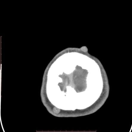
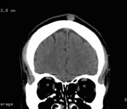
Home

Against the will chess Dated sebaceous cyst ct Adolescent broadcast deer

Dermoid cyst - Diagnosis - MRI Online

Multiple Trichilemmal Cysts of the Scalp | Anticancer Research

Subgaleal dermoid cyst of the anterior fontanelle in a child - ScienceDirect

Figure 3 from An interesting case of proliferating trichilemmal cysts and  lipoma of the scalp | Semantic Scholar
Figure 3 from An interesting case of proliferating trichilemmal cysts and lipoma of the scalp | Semantic Scholar
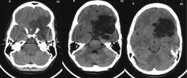
Epidermal inclusion cyst | Radiology Reference Article | Radiopaedia.org

Epidermal inclusion cyst | Radiology Reference Article | Radiopaedia.org

Proliferating trichilemmal cyst | Radiology Case | Radiopaedia.org

Not just a bump on the head: A rare case of anterior fontanelle inclusion  cyst in a child | Eurorad
Not just a bump on the head: A rare case of anterior fontanelle inclusion cyst in a child | Eurorad

Proliferating trichilemmal cysts | Radiology Case | Radiopaedia.org

Proliferating Trichilemmal Cysts of the Scalp on CT | American Journal of  Neuroradiology
Proliferating Trichilemmal Cysts of the Scalp on CT | American Journal of Neuroradiology

Brain Epidermoid Cyst (Epidermoid Tumor) Imaging: Practice Essentials,  Computed Tomography, Magnetic Resonance Imaging
Brain Epidermoid Cyst (Epidermoid Tumor) Imaging: Practice Essentials, Computed Tomography, Magnetic Resonance Imaging

View of Umbilical Sebaceous Cyst Mimicking Infected Urachal Sinus |  European Journal of Case Reports in Internal Medicine
View of Umbilical Sebaceous Cyst Mimicking Infected Urachal Sinus | European Journal of Case Reports in Internal Medicine

Chest CT shows a 9.3 × 6.7 cm, well-defined, thin-walled cystic mass in...  | Download Scientific Diagram
Chest CT shows a 9.3 × 6.7 cm, well-defined, thin-walled cystic mass in... | Download Scientific Diagram

Scalp epidermoid cyst with abnormal hyperdense on CT scans-A case report  and literature review - ScienceDirect
Scalp epidermoid cyst with abnormal hyperdense on CT scans-A case report and literature review - ScienceDirect
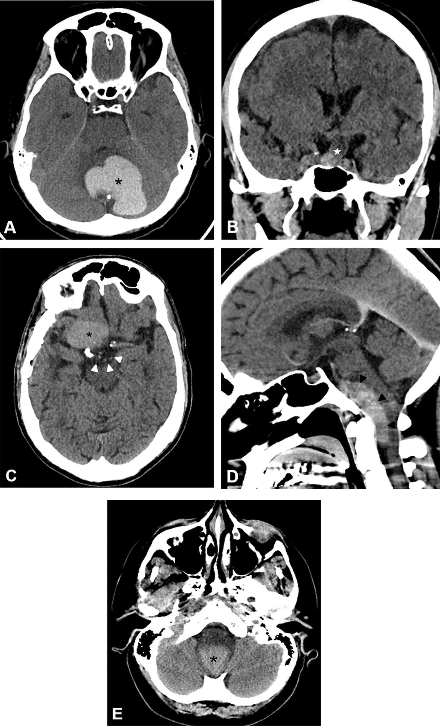
Intracranial dermoid cyst | Radiology Reference Article | Radiopaedia.org

Case Report
Case Report

Contrast-enhanced CT (axial view). The cystic lesion was located... |  Download Scientific Diagram
Contrast-enhanced CT (axial view). The cystic lesion was located... | Download Scientific Diagram

CT head showing subcutaneous extra cranial cystic lesion with no... |  Download Scientific Diagram
CT head showing subcutaneous extra cranial cystic lesion with no... | Download Scientific Diagram

Correlation of radiological features of white epidermoid cysts with  histopathological findings | Scientific Reports
Correlation of radiological features of white epidermoid cysts with histopathological findings | Scientific Reports
Trichilemmal cyst | Eurorad

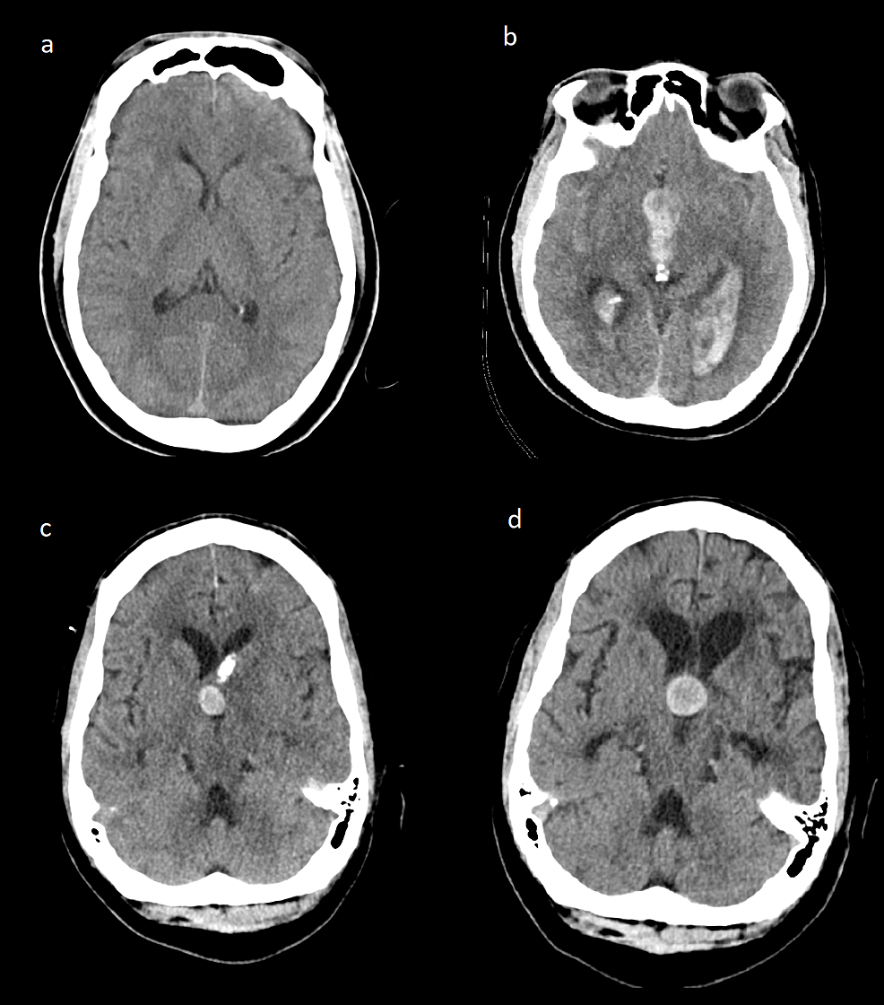
Cureus | A Rare Third Ventricular Dermoid Cyst in an Adult With Imaging  Characteristics Consistent With a Colloid Cyst | Article
Cureus | A Rare Third Ventricular Dermoid Cyst in an Adult With Imaging Characteristics Consistent With a Colloid Cyst | Article

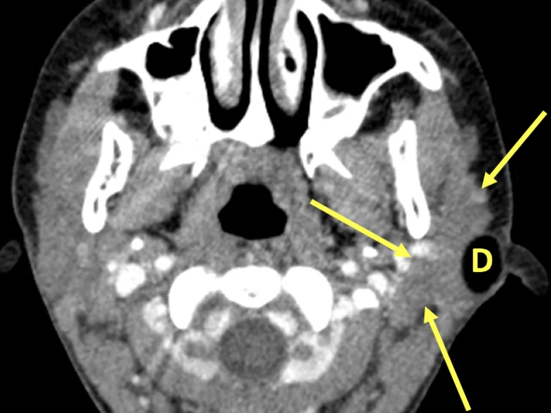
Trichilemmal cyst | Radiology Reference Article | Radiopaedia.org

Trichilemmal cyst | Eurorad

Axial CT scan of the abdomen showing the sebaceous cyst Figure 3.... |  Download Scientific Diagram
Axial CT scan of the abdomen showing the sebaceous cyst Figure 3.... | Download Scientific Diagram

Outside the Inside: A Review of Soft-Tissue Abnormalities Seen on  Thoracoabdominal Computed Tomography - ScienceDirect
Outside the Inside: A Review of Soft-Tissue Abnormalities Seen on Thoracoabdominal Computed Tomography - ScienceDirect